The Role of Precision Imaging in Modern Podiatric Care
Modern podiatry has moved far beyond the limitations of basic X-rays. While traditional radiography remains a standard tool for assessing bone alignment, advanced diagnostic imaging such as magnetic resonance imaging (MRI), computed tomography (CT), and musculoskeletal ultrasound allows clinicians to visualize soft tissue, joints, and complex internal structures with exceptional clarity.
High-quality imaging is essential for addressing intricate foot and ankle pathologies that standard exams often miss. By pinpointing conditions like subtle stress fractures, ligamentous tears, or inflammatory soft tissue damage early, specialists can move away from generalized treatments toward targeted, precision-based interventions. This transition often helps patients bypass unnecessary exploratory procedures in favor of faster, recovery-focused care.
At advancedfootcareil.com, our clinics in NW Chicago and South Florida prioritize this commitment to evidence-based medicine. We utilize these sophisticated tools to develop customized rehabilitation plans that account for the unique biomechanics of every patient. By integrating diagnostic precision into our standard protocols, we ensure that every treatment decision is grounded in an accurate, data-driven understanding of the underlying injury.
Essential Diagnostic Imaging Modalities for Foot and Ankle
 Modern podiatry relies on a sophisticated suite of diagnostic tools to move beyond simple surface examinations. Advanced diagnostic imaging provides clinicians with high-precision visuals of internal structures, which is essential for identifying the root cause of complex foot and ankle pain. By utilizing tools like MRI, CT scans, and ultrasound, specialists can accurately distinguish between overlapping symptoms to detect ligament tears, soft tissue injuries, and bone abnormalities that traditional X-rays might overlook. At advancedfootcareil.com, we leverage these advanced insights to develop personalized, minimally invasive treatment plans, effectively reducing the risk of misdiagnosis and unnecessary procedures.
What are the primary benefits of advanced diagnostic imaging in podiatry?
The primary benefit of this technology is the ability to visualize deep tissue structures that are invisible to the naked eye. While weight-bearing X-rays remain the foundation for assessing bone structure and alignment, advanced modalities offer greater specificity. MRI is widely considered the gold standard for complex soft tissue and bone marrow assessment, as it captures detailed images of tendons, ligaments, and cartilage without ionizing radiation. Unlike standard static imaging, diagnostic ultrasound provides the unique advantage of real-time, dynamic assessment, allowing doctors to observe tendons and muscles in motion during patient activity.
What are three common pathologies of the ankle and foot?
Three common pathologies that frequently affect the foot and ankle are bunions, plantar fasciitis, and Achilles tendon disorders. Bunions involve a structural deformity of the joint at the base of the big toe, while plantar fasciitis results from inflammation of the tissue along the bottom of the foot. Achilles tendon disorders involve injury to the large tendon connecting the calf to the heel. For complex cases involving these conditions, CT scans are invaluable for three-dimensional bone reconstruction, providing the precise geometric data necessary for planning intricate surgeries. While other clinics may rely solely on traditional methods, our approach at advancedfootcareil.com uses these imaging insights to ensure smaller incisions and accelerated recovery timelines.
| Modality | Primary Use | Key Advantage |
|---|---|---|
| X-Ray | Bone structure | Weight-bearing alignment |
| MRI | Soft tissue | High-detail resolution |
| Ultrasound | Dynamic motion | Real-time assessment |
| CT Scan | Bone geometry | 3D reconstruction |
Ultrasound and MRI: Choosing the Right Diagnostic Tool
 When is an MRI preferred over an ultrasound for evaluating foot and ankle pain? While ultrasound is a highly effective, cost-efficient tool for assessing superficial ligament injuries such as the ATFL and CFL, MRI remains the gold standard for evaluating deeper, more complex internal structures. MRI is specifically preferred when there is a clinical suspicion of syndesmotic injuries, which are often more challenging to visualize through ultrasound. Additionally, MRI is indispensable for the comprehensive assessment of bone marrow abnormalities and occult fractures that may not be clearly identified via ultrasound. Because these modalities offer complementary diagnostic strengths, a surgeon will often choose MRI when a clear, deep-tissue structural diagnosis is required for complex foot and ankle pain. Ultimately, the selection of imaging depends on whether the suspected injury is located in accessible surface tissues or requires the superior deep-tissue penetration and high-resolution imaging that MRI provides.
What types of conditions can be diagnosed using a foot or ankle ultrasound? A foot and ankle ultrasound is a versatile, non-invasive diagnostic tool that provides real-time, high-resolution imaging of soft tissues. It is highly effective for identifying musculoskeletal issues such as tendonitis, particularly Achilles tendonitis, and common conditions like plantar fasciitis. Beyond soft tissue injuries, ultrasound allows podiatrists to accurately diagnose ligament tears, bursitis, nerve compressions, and joint abnormalities. Additionally, this technology can visualize ganglion cysts, degenerative arthritis, and joint effusions with remarkable clarity. By offering a dynamic look at these internal structures, ultrasound plays a crucial role in both precise diagnosis and the guided delivery of therapeutic injections.
Comparing sensitivity and specificity in musculoskeletal imaging reveals distinct clinical advantages for each modality. For plantar plate injuries, ultrasound shows a high pooled sensitivity of 95 percent, making it an excellent screening tool for ruling out pathology. Conversely, MRI provides superior specificity at 83 percent, offering reliable confirmation when the extent of an injury must be mapped for surgical planning. At advancedfootcareil.com, our approach integrates these modalities to avoid minimally invasive podiatry guesswork. By utilizing the dynamic capabilities of ultrasound and the structural depth of MRI, we ensure that every treatment plan is grounded in precise, evidence-based imaging.
Diagnostic Precision for Surgical Planning and Recovery
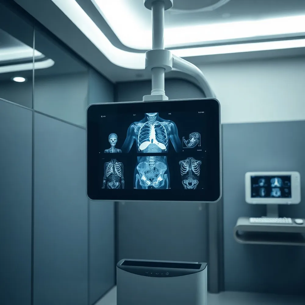 Advanced imaging is a foundational component of minimally invasive podiatry because it allows surgeons to visualize deep anatomical structures before a single incision is made. By employing high-resolution modalities like MRI and CT scans, practitioners can pinpoint the exact location of pathology, such as stress fractures or ligamentous tears, which ensures that surgical interventions are both targeted and effective. This level of precision stands in contrast to legacy approaches that often relied on broader, less specific examinations that could lead to unnecessary exploratory procedures.
Why is an MRI often necessary for diagnosing foot and ankle symptoms? An MRI is frequently necessary because it provides superior, highly detailed imaging of soft tissues, including ligaments, tendons, and cartilage that are often invisible on standard X-rays. While X-rays are excellent for identifying bone fractures or gross deformities, they cannot capture the nuances of complex injuries like chronic tendonitis, ligament tears, or subtle joint impingements. By utilizing advanced magnetic resonance technology, podiatrists can achieve a precise diagnosis for persistent pain that does not respond to initial conservative therapies. This diagnostic clarity is essential for developing effective, minimally invasive treatment plans tailored to the patient’s specific anatomical needs. Ultimately, incorporating MRI technology ensures an accurate assessment, allowing for earlier intervention and better long-term outcomes for complex foot and ankle conditions.
Beyond initial diagnosis, advanced imaging provides an objective timeline for recovery. Specialists use these tools to track healing progress, ensuring that hardware placement and tissue repair are proceeding as expected. At advancedfootcareil.com, this data-driven approach allows for the adjustment of rehabilitation plans in real time. Unlike facilities that may rely on subjective patient feedback alone, this objective monitoring ensures that any variance in recovery is identified immediately, helping patients achieve an accelerated recovery timeline while minimizing the risk of long-term complications.
Integrating Future Technology in Podiatric Diagnostic Protocols
Modern podiatry relies on digital radiography to capture high-resolution images while offering a significant reduction in radiation exposure. At advancedfootcareil.com, we employ these digital systems to provide immediate diagnostic clarity, allowing our specialists to manipulate contrast and zoom levels for a more comprehensive assessment. Unlike traditional film-based systems, this technology facilitates seamless integration into electronic health records, ensuring that follow-up comparisons are both accurate and efficient.
How do modern diagnostic imaging protocols guide the treatment of ankle injuries?
Diagnostic protocols such as the Ottawa Ankle Rules serve as essential decision-making tools to determine when radiographs are necessary, effectively reducing unnecessary radiation exposure. By assessing specific points of tenderness and a patient’s ability to bear weight, clinicians can efficiently rule out significant fractures and focus resources where they are truly needed. For complex cases where standard X-rays are insufficient, advanced imaging like MRI or CT scans allows for a high-resolution evaluation of ligamentous damage. This precise layering ensures treatment plans at advancedfootcareil.com are tailored to the individual, facilitating minimally invasive surgical techniques or targeted conservative therapies.
What are the latest technological advancements in podiatric diagnostics?
The field is rapidly evolving through the integration of sophisticated imaging and biomechanical tools. Beyond standard diagnostics, 3D foot scanning provides precise anatomical data that significantly improves the customization of orthotic devices to rectify biomechanical imbalances. Furthermore, digital gait analysis captures slow-motion biomechanical patterns, enabling a deeper understanding of how movement influences foot pathology. These advancements work in tandem with the minimally invasive podiatry techniques practiced at advancedfootcareil.com to streamline patient care, reduce recovery times, and enhance long-term functional outcomes.
Commitment to Excellence in Foot and Ankle Diagnostics
Selecting the right specialist is as important as the technology itself. Because musculoskeletal ultrasound and other imaging modalities are highly operator-dependent, the accuracy of a diagnosis rests on the clinician's ability to interpret complex anatomical layers. At advancedfootcareil.com, we prioritize this expertise by pairing advanced diagnostic imaging with specialized surgical knowledge.
Precise imaging serves as the backbone of evidence-based medicine. By utilizing high-resolution tools to confirm the extent of a pathology before intervention, surgeons can avoid unnecessary exploratory procedures and target the specific source of pain. This commitment to diagnostic excellence ensures that every treatment plan is tailored to the patient, resulting in faster recovery times and better long-term functional outcomes.